+91 9645 600 032 | +91 9895 780 909
Mon to Sat 8.00 am to 6.00 pm
Improve your vision with our LASIK and Cornea treatments.
LASIK, or laser-assisted in situ keratomileuses, swiftly reshapes the cornea, enhancing light focus and improving vision.
Our eye surgeons tailor LASIK to reduce your dependency on glasses or contacts. The entire procedure takes just 15-20 minutes and is pain-free. Additionally, the LASIK procedure ensures rapid healing, improving vision clarity within days.
Experience the transformative power of LASIK and Cornea treatments for a clearer, brighter vision.
We deliver the following Cornea treatments:
-
SCHIRMER’S TEST
-
SOFT LENS / TORIC LENS / RUP
-
TOPOGRAPHY / SPECULAR MICROSCOPY
-
C3R / T CAT
-
ABERROMETRY / LIPISCAN / LIPIFLOW
-
ICL IMPLANTATION
-
LASIK & PRK TREATMENT
-
PTK
Corneal diseases
Corneal diseases can cause clouding and distortion of vision, and eventually blindness. There are many types of corneal diseases like infections due to contact lenses, dry eye, abrasions from trauma, and inflammations. Other conditions include keratoconus, pterygium etc.
The layers of the cornea
The epithelium is layer of cells that cover the surface of the cornea. It is only about 5-6 cell layers thick and quickly regenerates when the cornea is injured. If the injury penetrates more deeply into the cornea, it may leave a scar. Scars leave opaque areas, causing the corneal to lose its clarity and luster.
Boman's membrane lies just beneath the epithelium. Because this layer is very tough and difficult to penetrate, it protects the cornea from injury.
Descemet's membrane lies between the stroma and the endothelium. The endothelium is just underneath Descemet's and is only one cell layer thick. This layer pumps water from the cornea, keeping it clear. If damaged or disease, these cells will not regenerate.
Tiny vessels at the outermost edge of the cornea provide nourishment, along with the aqueous and tear film.
What is Keratoconus?
Keratoconus is a frequently seen corneal disease, occurring in about 1 in 1000 people, which typically starts after the age of 10 yrs. The hundreds of filaments of collagen layers in normal cornea are linked to each other by cross linking, giving it an enormous strength. If these collagen cross-links are lost, as it happens in Keratoconus, there will be a progressive corneal thinning and stretching. This often occurs in both the eyes. The cornea bulges forward into an irregular cone shape. This causes distortion of the image, which cannot be corrected with glasses. The eye develops irregular astigmatism (cylindrical errors) and myopia [shortsightedness] and the vision would become blurred.
Who gets Keratoconus?
Risk factors include eye rubbing, family history of keratoconus, genetic predisposition, certain systemic disorders such as Down’s syndrome, ocular allergy, connective tissue disease etc. Diabetics won’t develop Keratoconus because of natural crosslinking from high glucose and UV light. Affects men and women in equal proportions and is bilateral in 90% of cases.
What are the symptoms?
At early stages, the person feels the need for frequent change of spectacle correction. As the disease progresses, the vision deteriorates. Visual acuity becomes impaired at all distances, and night vision is sometimes quite poor. Keratoconus can cause substantial distortion of vision, with multiple images, streaking, sensitivity to light, eye strain from squinting in order to read, & itching in the eye.
How it is diagnosed?
This is usually done by an Ophthalmologist with a detailed eye examination including retinoscopy, keratometry, slit-lamp examination etc. Diagnosing early keratoconus can be tricky, since mild disease often does not show any signs on slit-lamp examination. Streak retinospcopy can pick up early Keratoconus.
At Sreekanth eye care & research centre Netralaya a very sensitive instrument called the ‘Pentacam HR’ is used for early diagnosis. This is an automated instrument working on Shiemflug principal, and can pick up extremely early Keratocouns, that often starts in the back of the cornea.
How Keratoconus is treated?
Treatment of mild to moderate keratoconus is done temporarily by Contact lenses, which can be the regular RGP or specific and specialised, like Rose K lenses and mini scleral contact lenses depending upon the severity of the illness. The new modality of treatment is Corneal Collagen Crosslinking with Riboflavin (C3-R) stabilises the cornea from further deteriorating. In severe stages the person has to undergo corneal grafting surgery.
A Few About LASIK
Make an Appointment A Few About Cataract Refractive errors occur when an eye cannot clearly focus the image on the retina, resulting in blurred vision. This in many cases can subsequently lead to a permanent un-correctable disability. What Causes Cataract? How does Cataract Affect Vision? How is Cataract Detected? When must a Cataract be Removed? How is Cataract Surgery Performed? What are the types of Artificial Lens Implants? Monofocal and multifocal lens implants are available. With multifocal implants, you may not need eyeglasses for reading. Your eye surgeon will discuss with you and recommend the type of implant that is most suitable for you. A Few About LASIK LASIK stands of “Laser Assisted In Situ Keratomileusis”. Performed first in 1990 in the United States, LASIK boasts to have one of the highest patient satisfaction rates among medical procedures worldwide today. LASIK involves extremely precise and accurate reshaping and sculpting of the cornea to a predetermined curvature using an Excimer LASER, so as to focus the image on the retina. It is also called as LASER vision correction.
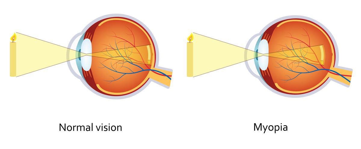
Myopia ( Short sightedness)
Is a condition where image is focused in front of the retina either because the eye is too long or its focusing power is too strong. The distance objects appear blurred.

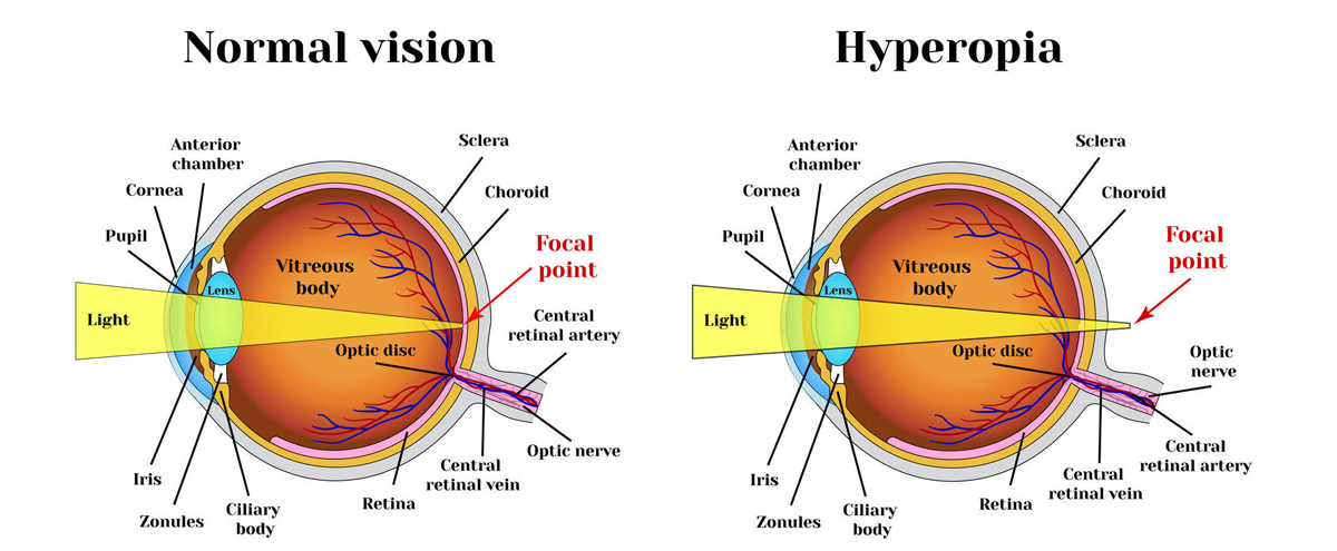
Hyperopia (Long sightedness)
Here the light rays reach the retina even before it gets focussed. The eye is too short or its focusing power is weak. Both distance & near objects are blurred and initially blurring may be intermittent. Eye fatigue often occurs and this is very common especially in children.

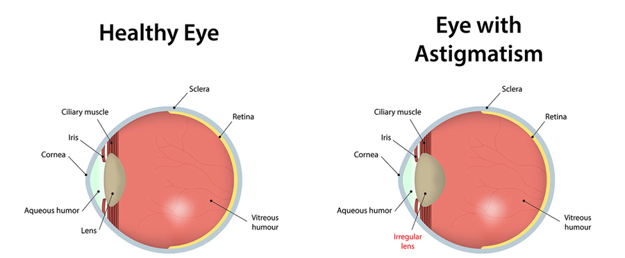
Astigmatism
Here the eye does not focus light evenly onto the retina due to irregular shape of the cornea.

Presbyopia
Is a normal ageing process. The ciliary body of the eye is unable to work effectively, and do not focus the image of near objects sharply on the retina. This results in a need to wear near glasses for reading and near work. Presbyopia may be associated with any of the above three conditions


